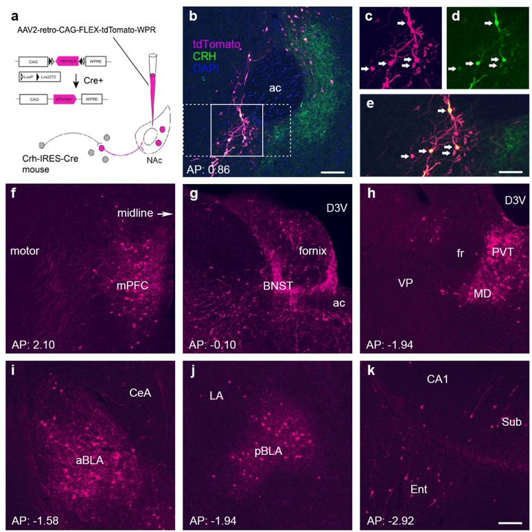
Figure 1. The major CRH-expressing afferents to the nucleus accumbens originate in the paraventricular nucleus of the thalamus, the bed nucleus of the stria terminalis, the medial prefrontal cortex and the basolateral amygdala.
(a) Schematic of the retrograde-labeling virus and the location of the injection site. Specifically, a Cre-driven adeno-associated virus (AAV; AAV2-retro-CAG-FLEX-tdTomato-WPRE) was injected into the left hemisphere NAc in CRH-IRES-Cre mice. (b–e) Endogenous CRH+ cells in the nucleus accumbens (NAc) were infected by the rAAV2-retro. (b) Low magnification image from a representative injection site, the NAc in the left hemisphere (n = 5 mice). The labeling of cell bodies and fiber terminals in the NAc is apparent. The tdTomato reporter for the virus is shown in magenta, whereas immunostaining to confirm local CRH+ neurons is shown in green. The section was counterstained with DAPI (blue). Framed areas in b were magnified in c–e to demonstrate virus-labeled CRH cells (c), antibody-immunolabeled CRH cells (d), and the co-expression of virus label and endogenous CRH (e). (f–k) Anterior-posterior series of panels demonstrating significant sources of retrogradely labeled CRH+ NAc afferents. Labeled cells and fibers were primarily found in the ipsilateral (left) hemisphere. (f) The medial prefrontal cortex (mPFC) including prelimbic and infralimbic regions contributed 5.75% of CRH+ projections to the NAc. (g) 9.27% of CRH+ inputs originated from the bed nucleus of the stria terminalis (BNST). (h) Thalamic nuclei were a major source of CRH projections to the NAc, contributing over a third (34%). Among them, the paraventricular nucleus (PVT) accounted for a third of all thalamic NAc-projecting cells (9.40% of the total). (i–j) 8.57% of CRH+ projections came from amygdala nuclei, and over half from the BLA. (k) CRH+ inputs arise from the subiculum and the entorhinal cortex. ac: anterior commissure; arrows in c-e indicate dual-labeled cell bodies of CRH cells. ac = anterior commissure; aBLA = anterior basolateral amygdala; BNST = bed nucleus of the stria terminalis; CeA = central amygdala; D3V = dorsal third ventricle; Ent = entorhinal cortex; LA = lateral amygdala; mPFC = medial prefrontal cortex; pBLA = posterior basolateral amygdala; PVT = paraventricular nucleus of the thalamus; Sub = subiculum. Bar = 100 µm in b, 63 µm in c–e and 100 µm for f–k.

