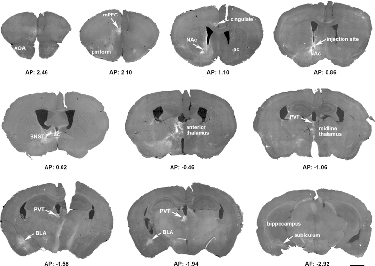
Figure 2. Overview of a representative mouse brain, highlighting the sparse, selective origins of CRH+ NAC-directed inputs.
A Cre-driven adeno-associated virus (AAV; AAV2-retro-CAG-FLEX-tdTomato-WPRE) was injected into the left hemisphere nucleus accumbens (NAc) in a CRH-IRES-Cre mouse. The 15 coronal sections (30 µm) demonstrate the location of reporter-labeled cells and fibers through the rostro-caudal axis of the brain. Sections were obtained from 2.8 mm to −4.72 mm AP relative to the Bregma. ac = anterior commissure; AOA = anterior olfactory are; BLA = basolateral amygdala; BNST = bed nucleus of the stria terminalis; mPFC = medial prefrontal cortex; PVT = paraventricular nucleus of the thalamus. Bar = 1 mm.

