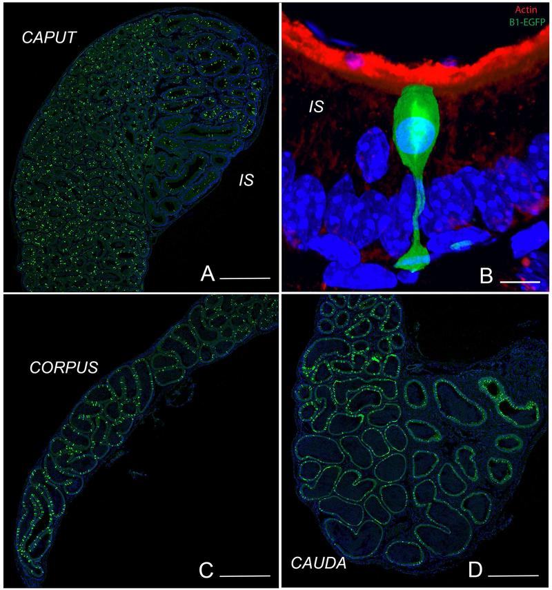
Figure 1). Visualization of NCs and CCs in the epididymis of transgenic mice expressing EGFP under the control of the promoter of the V-ATPase B1 subunit (B1-EGFP).
EGFP+ NCs (green) are located in the IS, and EGFP+ CCs (green) are located in the caput (A), corpus (C) and cauda (D) regions. B) In the IS, NCs have a “champagne glass” appearance and their nuclei are located in the apical region of the epithelium, compared to adjacent PCs. A dense network of filamentous actin is seen in the apical stereocilia of PCs (labeled in red using phalloidin). Nuclei are labeled in blue using DAPI. Bars: A, C, D = 500 μm, B = 5 μm.

