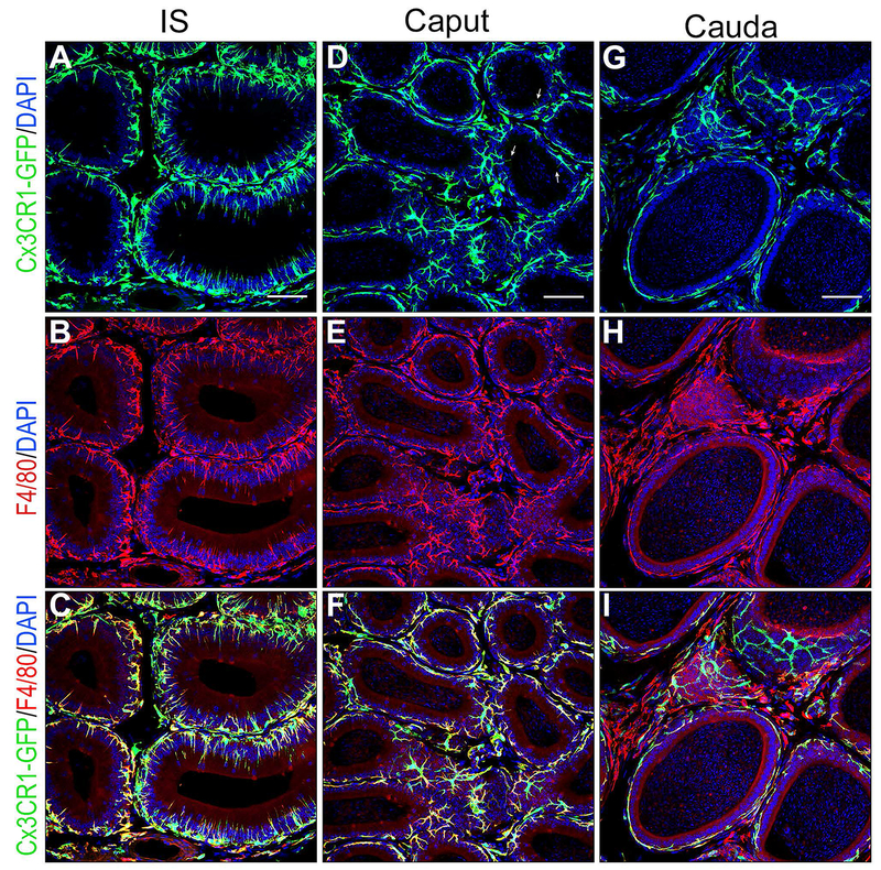
Figure 6). Confocal microscopy showing a dense network of mononuclear phagocytes in all epididymal segments.

Epididymis of CX3CR1-EGFP transgenic mice was labeled for the macrophage marker F4/80 (red). A,B,C) In the IS, most CX3CR1 positive MPs (green) are also labeled for F4/80 identifying them as macrophages. The majority of macrophages are in close proximity with the epithelium and show numerous intraepithelial projections extending toward the lumen. D,E,F) In the caput, most CX3CR1 positive MPs (green) are also labeled for F4/80 identifying them as macrophages. These macrophages are in close proximity with the epithelium but only a few rare cells now send intraluminal projections towards the lumen (arrows in D). A significant number of cells positive for both CX3CR1 and F4/80 are located in the interstitium. G, H, I) In the cauda, cells positive for CX3CR1 and F4/80 are located next to the epithelium but they do not extend intraepithelial projections. In the interstitium, a mixed population of cells double positive for CX3CR1 and F4/80, and cells positive for F4/80 but negative for CX3CR1 are detected. Nuclei are labeled in blue with DAPI.
