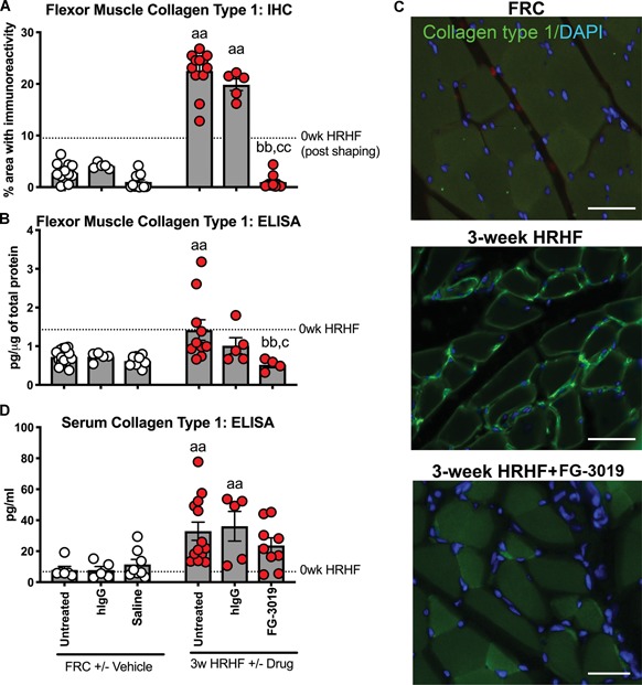
Figure 3.

Collagen type 1 in flexor digitorum muscle and serum. (A) Percent area with collagen type 1 immunoreactivity (green) and 4′,6‐diamidino‐2‐phenylindole (DAPI) nuclear stain (blue) in cross‐sectionally cut muscles of food‐restricted control (FRC), 3‐week untreated high repetition high force (HRHF) and HRHF + FG‐3019‐treated rats. (B) Enzyme‐linked immunosorbent assay (ELISA) detected levels in muscles. (C) Collagen type 1 serum levels, tested using ELISA. aa: p < 0.01, compared with matched FRC group; bb: p < 0.01, compared with HRHF rats; c: p < 0.05, and cc: p < 0.01, compared with HRHF + hIgG rats. Scale bars = 50 μm. The 0‐week HRHF rat results from Supplementary Table 1 are indicated with dotted lines [Color figure can be viewed at wileyonlinelibrary.com]
