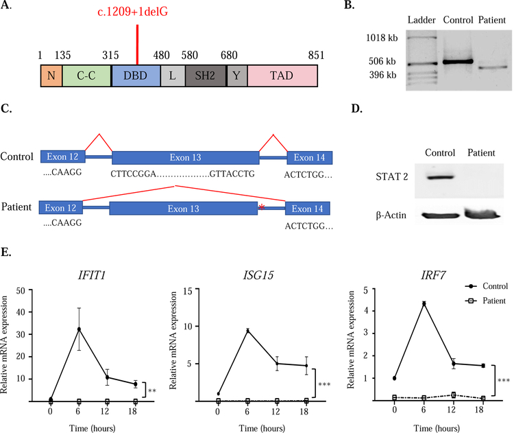
Figure I. The STAT2 homozygous splice donor variant mutation abrogates protein expression.
A. Linear map of STAT2 with the patient’s variant (c.1209+1delG)indicated in red. B. RT-PCR amplification of STAT2 mRNA encompassing the patient mutation site. C. Sanger sequencing of cDNA in the region surrounding the mutation in patient and control. Asterisk indicates mutation location. D. STAT2 protein expression in lysates from BLCLs from patient and control. E. Quantitative PCR analysis of IFIT1, ISG15 and IRF7 mRNA at baseline and after IFN-α stimulation of EBV-transformed BLCLs from the patient and controls (n=3). Values were normalized to GAPDH and expressed relative to the mean of unstimulated controls. Symbols and bars in E represent mean and SEM. ** p<0.01, *** p<0.001. Similar results were obtained in two independent experiments in B, D and E.

