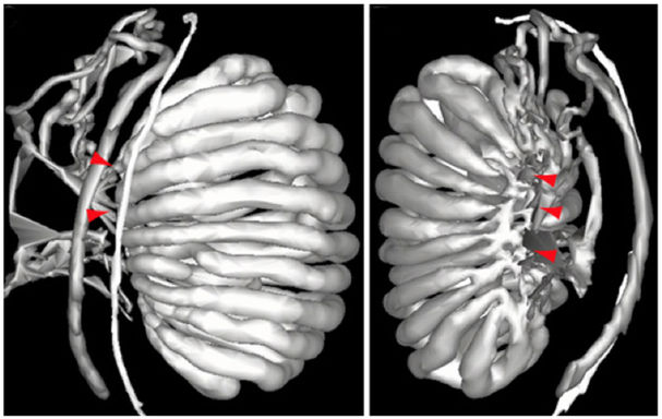
Figure 2.
Two three-dimensional models from an E15.5 mouse embryo showing the connectivity between the testis cords and the mesonephric tubules. Connections are labeled with red arrowheads. It is at these points that a plexus forms with perforations, which then contracts and narrows to form the rete testis on the dorsomedial aspect of the testis. The images were kindly provided by Dr. Peter Koopman, with the left image (lateral view) being published (Combes et al., 2009) and reproduced by kind permission of Wiley Press.

