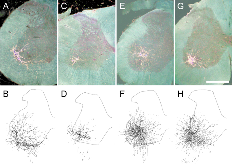
Figure 3.
Darkfield digital micrographs of transverse hemisections through the lumbar spinal cords and computer-generated reconstructions BHRP-labeled somata and processes of an untreated animal (A,B), and saporin-injected animals with either no further treatment (C,D) or given ad lib exercise (E,F), and an intact animal given ad lib exercise (G,H) after BHRP injection into the left vastus lateralis muscle. Computer-generated composites of BHRP labeling were drawn at 480 μm intervals through the entire rostrocaudal extent of the quadriceps motor pool; these composites were selected because they are representative of their respective group average dendritic lengths. Scale bar = 500 µm.

