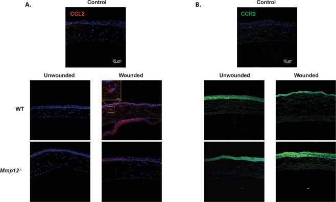
Figure 2.
Expression patterns of CCL2 and CCR2 in unwounded and wounded corneas of WT and MMP12 KO mice. (A) Immunofluorescence of CCL2 chemokine and (B) its receptor CCR2 in unwounded and chemically wounded (2-days after injury) WT and Mmp12−/− mouse corneas. Control images represent mouse corneas stained with secondary antibody only and without primary antibody. Nuclei were visualized by staining with DAPI (blue). Scale bars: 50 µm. A magnified image of a wounded WT cornea shows perinuclear expression of CCL2 (orange box). CCL2 staining was visualized in epithelial, stromal, and endothelial layers of wounded WT and Mmp12−/− corneas. CCR2 staining was visualized in epithelial and stromal layers of unwounded and wounded WT and Mmp12−/− corneas.

