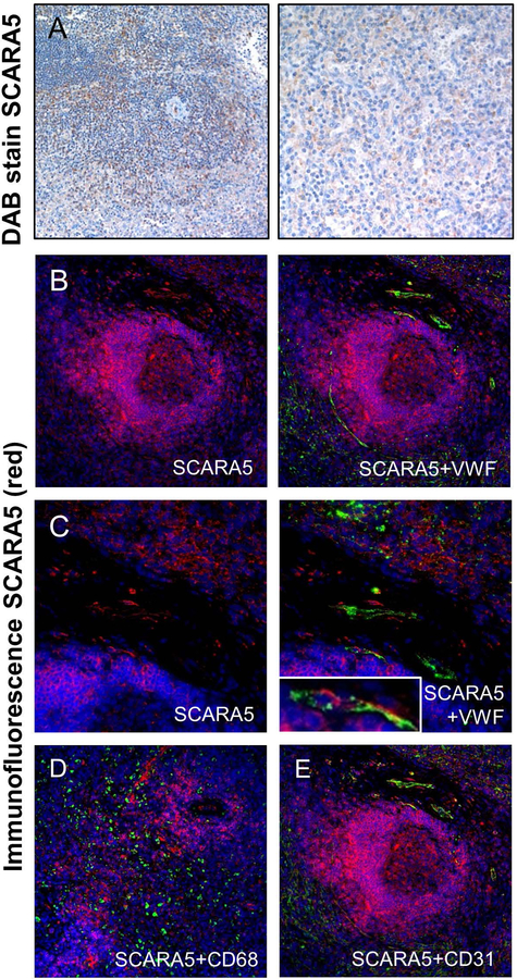
Figure 6. SCARA5 protein expression in the human spleen.
(A and B) DAB stain of SCARA5 expression in the human spleen (brown). (C) Immunofluorescent stain of SCARA5 in the spleen (red) (20×). (D) VWF (green) and SCARA5 (red) immunofluorescent imaging in the spleen (20×). (C) Immunofluorescent staining of SCARA5 (red) in the spleen (40×). (D) VWF (green) and SCARA5 (red) immunofluorescent imaging in the spleen (40×). (E) SCARA5 (red) and CD68 (green) immunofluorescent imaging in the spleen (20×). (D) SCARA5 (red) and CD31 (green) immunofluorescent imaging in the spleen (20×). For all images, DAPI = blue.

