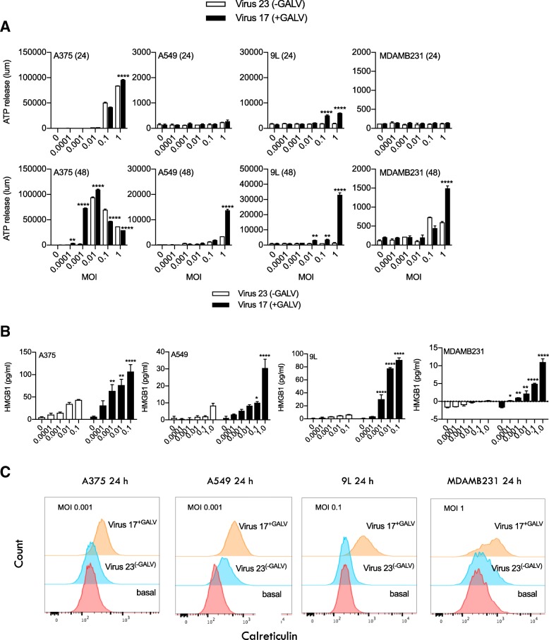
Fig. 3.
Markers of immunogenic cell death in cells treated with either Virus 23 (expresses hGM-CSF) or Virus 17 (expresses hGM-CSF and + GALV-GP R-) in vitro. a Levels of ATP release measured by luminescence in a panel of cell lines treated at the indicated MOI at 24 h post infection and (a) at 48 h post infection observed in cell-free supernatants treated with Virus 23 (indicated by the clear bars) and Virus 17 (indicated by the solid bars). b ELISA measuring HMGB1 (pg/ml) levels in cell-free supernatants from cells treated for 48 h with MOI 0.0001–1. c Histogram showing the expression levels of surface calreticulin (CRT) in cells treated at indicated MOI 0.01 for 48 h. Data show un-permeabilized, viable cells stained with CRT and measured by FACS. Statistical differences between groups were determined by using two-way ANOVA, *p < 0.05, **p < 0.01, ***p < 0.001, ****p < 0.0001

