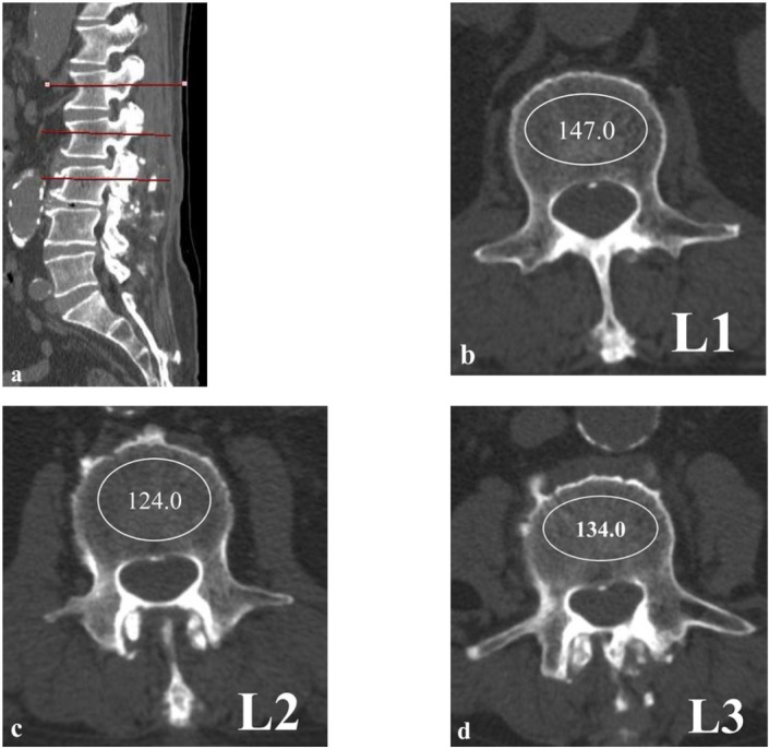
Figure 1.

Hounsfield Units measurement by drawing elliptical ROI on lumbar CT scan. The largest ROI is drawn excluding the cortical bone and vascular markings at mid-vertebral body from each vertebra.
(a) Sagittal image, (b) L1 axial image, (c) L2 axial image, (d) L3 axial image
