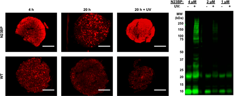
Figure 6.
Left: Spheroids were treated with 1 μM N23BP (top) or WT (bottom) affibody for a total of 4 h (left column) and 20 h (middle and right columns) and additionally irradiated with 365 nm light for 30 min after 3.5 h incubation (right column). These spheroids were sectioned at approximately the same depth and imaged for Rhodamine and GFP distribution. Micrographs show Rhodamine signal within each section normalized by the average GFP intensity. Scale bars are 200 μm. Right: Five spheroids were grown and irradiated for 1 h with the indicated N23BP affibody concentration in growth media. Affibody containing media was removed, and spheroids were lysed and loaded onto a PAGE gel and probed for affibody conjugates using an anti-T7 antibody. High molecular weight bands around the expected molecular weight of EGFR were observed only when the spheroid-affibody mixtures were irradiated, indicating photo-cross-linking.

