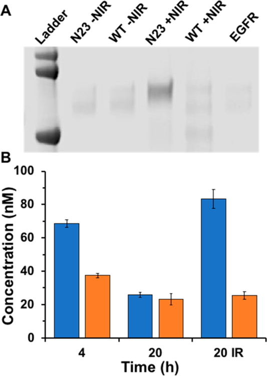
Figure 8.

(A) Denaturing polyacrylamide gel electrophoresis (SDS-PAGE) showing photo-cross-linking products of either N23BP or WT to EGFR, with or without irradiation at 980 nm. Note that only 980 nm irradiation of N23BP produced a photoproduct significantly different than free EGFR (right lane). Ladder proteins are (top to bottom) 100, 75, and 50 kDa. (B) Retention of fluorescently labeled affibodies (left, N23BP; right, WT) in 3D tumor spheroids grown from transfected 4T1 cells either induced with 15 μg/mL cumate. Concentrations of retained affibodies were calculated by comparing spheroid lysate fluorescence to standard curves prepared for each labeled affibody. “20 IR” designates that this sample was irradiated at 980 nm after 3 h incubation, then left to grow to a total of 20 h. Lanes without an IR designation were not irradiated.
