Figure 5:

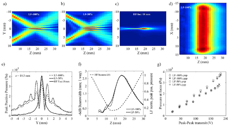
Cross-sections in the elevation plane of the transmit LF beam with: (a) 100%-BW excitation, (b) 50%-BW excitation and (c) transmit HF beam focusing at 18 mm showing the alignment of the center axes of the LF and HF component of the dual-frequency probe; (d) cross-section of the LF beam in the azimuthal plane showing the uniformity of the plane wave amplitude along the HF array length (23 mm); (e) HF and LF elevation beam profiles measured on transmit at LF focal distance; (f) LF transmit beam profiles on center axis superimposed on the variations of the HF beamwidth as a function of depth; (g) LF peak pressure as a function of transmit voltage amplitude.
