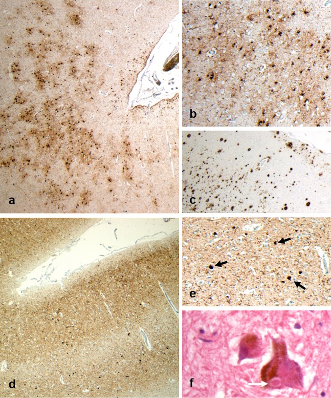
Fig. 2.
Representative images from Case 3, a former soccer player who first came to attention in his 60 s with mild cognitive impairment. There followed a steady progression in these symptoms, with exacerbations in the post-operative period, and with the development of visual hallucinations and motor signs leading to a clinical suspicion of Parkinson’s disease, or related disorder. After an 8-year course, the patient died in his 70 s. Staining for p-tau reveals patches of neuronal and astroglial immunoreactivity at the depths of cortical sulci (a), which on higher power are clustered around small cortical vessels (b; PHF1). Frequent Aβ plaques are also present (c; 6f3d). In addition, staining for alpha-synuclein reveals extensive cortical staining (d), with numerous cortical Lewy bodies present (e; black arrows). Sections of the midbrain also reveal scattered classical Lewy bodies (f; white arrow) within remaining pigmented neurons of the substantia nigra. Given the stereotypical clinical presentation and associated neuropathological features, an integrated diagnosis of dementia with Lewy bodies was made, with co-morbid CTE-NC noted

