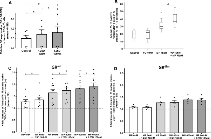
Fig. 1.
a GR protein expression normalized to glyceraldehyde 3-phosphate dehydrogenase (GAPDH) after incubation of human CD3+ T cells with 1,25D in vitro (24 h; PHA 0.5 µg/ml; n = 6 per group. Cell-based ELISA). Data are expressed as ratio of optical density (OD) 450 GR/GAPDH. b Apoptosis of human CD3+ T cells of healthy donors. Incubation with control (solvent: 0.1% DMSO), 1,25D, MP or 1,25D + MP in vitro (72 h; PHA 0.5 µg/ml; n = 7 per group; Annexin V/PI, flow cytometry). Apoptosis of murine splenic-derived CD3+ T cells from c BALB/c GRwt (wild type) or d BALB/c GRdim mice. Incubation with control (solvent: 0.1% DMSO), 1,25D, MP or 1,25D + MP in vitro (24 h; ConA 1.5 µg/ml; n = 9–10 (GRwt); n = 5 (GRdim); Annexin V/PI, flow cytometry). Apoptosis is depicted as increase over control (–; control: solvent 0.1% DMSO). In the control groups of each genotype, mean percentage of apoptotic murine T cells (SE) was as follows: BALB/c GRwt 46.1% (4.3) vs BALB/c GRdim 42.3% (3.6). GR glucocorticoid receptor, OD optical density, 1,25D 1,25(OH)2D3, MP methylprednisolone, SE standard error. Statistics: Wilcoxon signed-rank test: # < 0.05

