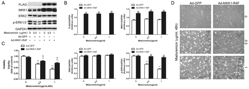
Fig. 3.
Ectopic expression of constitutively active MKK1 intervenes toxicity of maduramicin in H9c2 cells. H9c2 cells, infected with Ad-MKK1-R4F or Ad-GFP (as control), respectively, were exposure to maduramicin at indicated concentrations for 24 h (for Western blotting) or 48 h (for cell viability assay and cell proliferation analysis). A. Total cell lysates were subjected to Western blotting using indicated antibodies. B. Blots for indicated proteins were semi-quantified using NIH image J. C. Cell viability was detected by one solution assay. D. Cell images were taken with an Olympus inverted phase-contrast microscope equipped with the Quick Imaging system. For (A), representative blots were shown. The blots were probed for GAPDH as a loading control. Similar results were observed in at least three independent experiments. For (B), all data were expressed as means ± SE (n = 3). Statistical analysis was performed using two-way ANOVA followed by Bonferroni’s post-test. ** P < 0.01, difference with control group; ## P < 0.01, Ad-MKK1-R4F group versus Ad-GFP group. For (C), all data were expressed as means ± SE (n = 6). Statistical analysis was performed using two-way ANOVA followed by Bonferroni’s post-test. ** P < 0.01, difference with control group; ## P < 0.01, Ad-MKK1-R4F group versus Ad-GFP group. For (D), representative photos were shown. Similar results were observed in at least three independent experiments.

