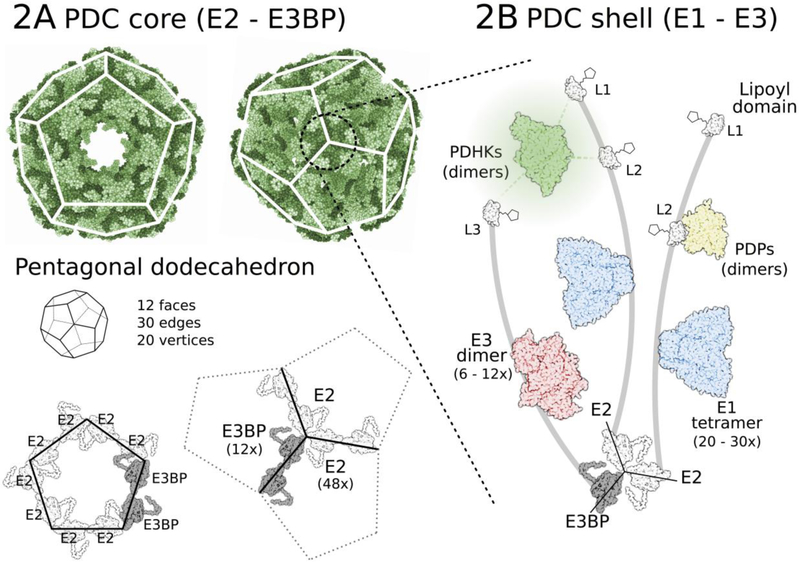
Figure 2: Pyruvate dehydrogenese complex structure (9.5 MDa).
(A) PDC core consisting of 48 E2 and 12 E3BP components is assembled into a pentagonal dodecahedron (3.5 MDa). (B) 20–30 E1 (α2β2 tetramer) components attached to the subunit binding domains of E2 and 6–12 E3 (α2 dimer) units on E3BP make up a plastic shell on the PDC core. Regulatory PDHKs (α2 dimers) and PDPs (αβ dimers) are also part of this shell, binding to lipoyl domains (mostly inner lipoyl L2 domains) on the flexible arm regions of E2 and E3BP. Such formation facilitates easy movement of individual enzymes and reaction intermediates over the complex.

