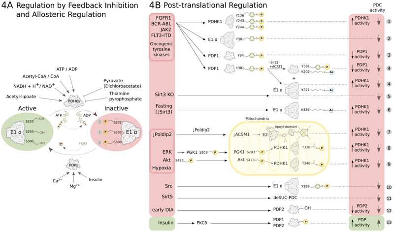
Figure 4: Regulation of PDC activity.
(A) PDC activity is regulated by reversible phosphorylation and dephosphorylation of its E1α subunit executed by PDHKs and PDPs. PDHKs perform inhibitory phosphorylation of three E1α serine residues (Ser 293 – site 1, Ser 300 – site 2, Ser 232 – site 3) and PDPs activate PDC by dephosphorylating these three sites. High PDC reaction endproduct/substrate ratios activate PDHKs by feedback inhibiton, while pyruvate or its synthetic analogue dichloroacetate inhibit PDHKs allostericaly (details in text). PDPs are activated by Ca2+ and Mg2+ ions, and insulin. (B) Post-translational regulation of PDC activity (details in text). (1)97, (2)100, (3)101, (4)102, (5)103, (6)104, (7)107, (8)98, (9)99, (10)105, (11)106, (12)130, (13)60.

