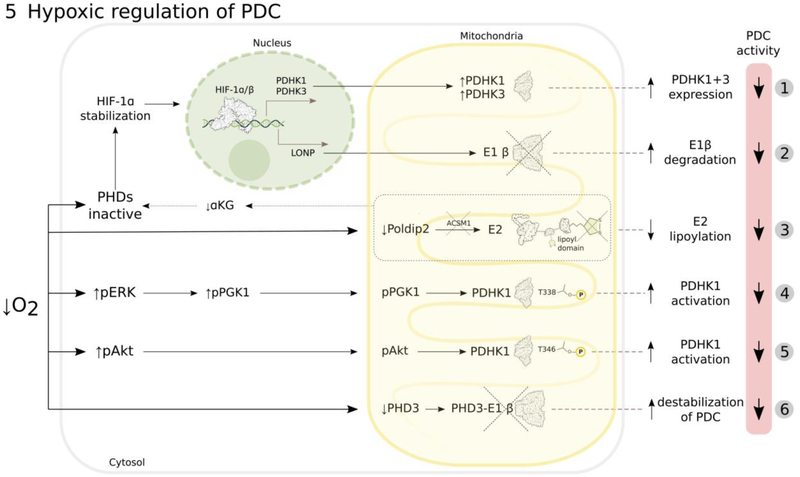
Figure 5: Mechanisms involved in regulation of PDC activity by tumor hypoxia.
Hypoxia decreases PDC activity (more details in text) by: HIF-1-mediated transcription of PDHK1 and PDHK3, which inhibit PDC by phosphorylation of E1α (1)15–17; HIF-1-induced transcription of LONP protease that degrades E1β (2)40; downregulation of Poldip2, resulting in increased degradation of the lipoic-acid activating enzyme ACSM1, which leads to decreased E2 lipoylation and PDC activity, accompanied by decreased α-ketoglutarate (αKG) levels that additionally inhibit prolyl-hydroxylases (PHDs) and stabilize HIF-1α (3)107; activating PDHK1 by post-translational phosphorylation of its threonine residues either by ERK-phosphorylated and mitochondrially-translocated phosphoglycerate kinase 1 (PGK1) (4)98 or by mitochondrially-translocated phosphorylated Akt (5)99; prolyl-hydroxylase PHD3 depletion that leads to PDC destabilization due to decreased PHD3-E1β interaction (6)41.

