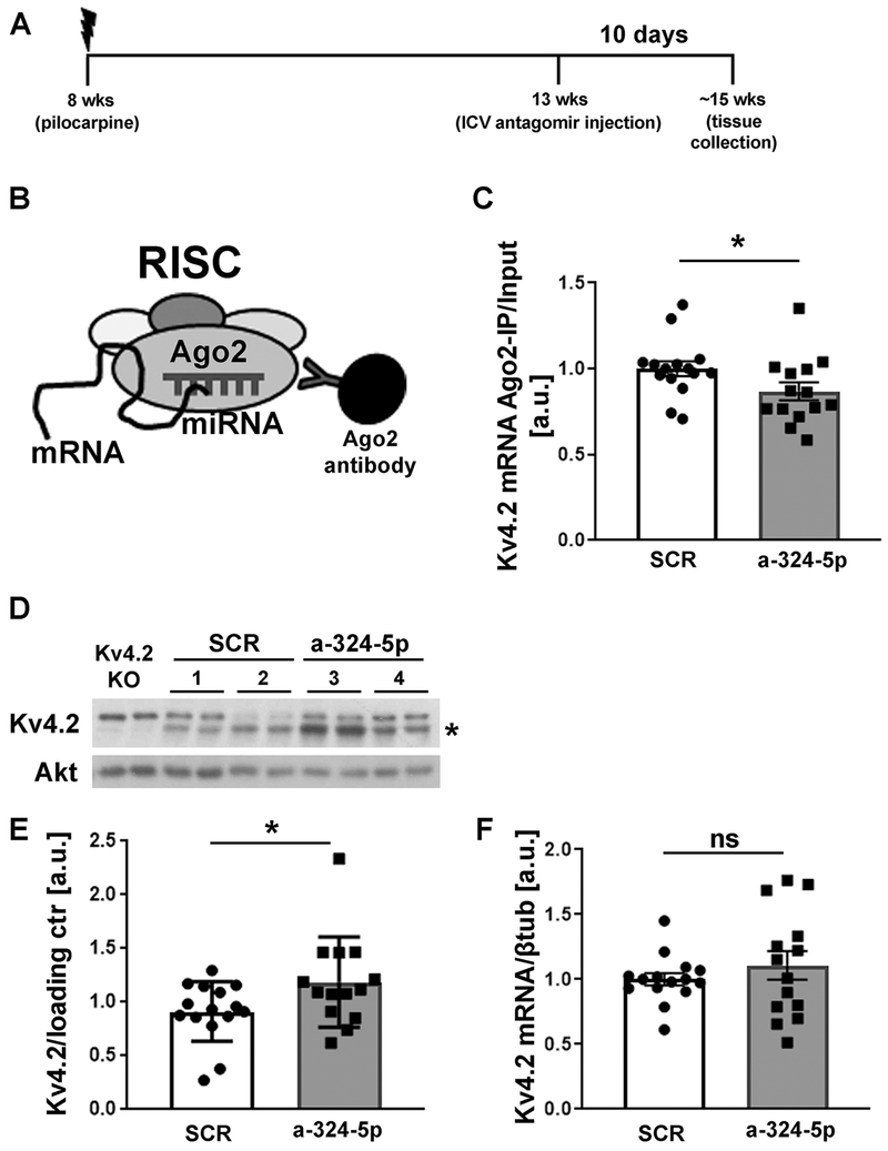
Figure 4: miR-324-5p antagomir reduces microRNA-induced silencing of Kv4.2 in the pilocarpine model.
(A) Timeline depicting age of the mice during pilocarpine injection, antagomir treatment and tissue collection. (B) Schematic of Ago2-specific immunoprecipitation to assess Kv4.2 mRNA levels in the RISC. (C) Reduced association of Kv4.2 mRNA with Ago2 was observed after miR-324-5p antagomir treatment compared to scrambled control (C: SCR: n=15, a-324-5p: n=14, unpaired one-tailed t-test, t(27)=1.940, *p=0.031). mRNA levels in Ago2-IPs were quantified by qRT-PCR and normalized to input levels. (D,E) Western blot analyses of hippocampal lysates show significantly increased Kv4.2 expression in chronically seizing mice 10 days after miR-324-5p antagomir treatment compared to scrambled control (SCR: n=15, a-324-5p: n=14, unpaired one-tailed Mann-Whitney test, *p=0.047). Example blot of 2 different mice per condition (#1-4, loaded in duplicates) shown in D, cumulative quantification shown in E. Kv4.2-specific signal was normalized to loading control (Akt or βTubulin) on the same blot. Asterisk indicates Kv4.2-specific band that is absent in lysates from Kv4.2 KO mice. (F) Kv4.2 mRNA levels (normalized to βtubulin) were not significantly changed after antagomir treatment (SCR: n=15, a-324-5p: n=14, unpaired one-tailed t-test, t(27)=0.916, p=0.184). Error bars represent SEM.

