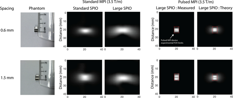
Fig. 10. Verification of pulsed MPI image resolution improvement using line phantoms in 2D imaging.

(Left) Line phantoms used; one is separated by 0.6 mm and the other by 1.5 mm. (Center) Standard MPI images line phantoms taken with our preclinical FFL system at 3.5 Tm−1 with the current standard MPI tracer: (VivoTrax™ ) and a monodisperse large core (27.4 nm) tracer. With standard MPI, the large core tracer shows more blurring than the standard tracer. (Right) Pulsed MPI data using the large core tracer. Our tabletop AWR in 2D mode has a limited FOV denoted by the dashed red lines. For visual comparison with the preclinical, standard MPI results, we display the pulsed MPI images in the larger total FOV by filling in the unsampled regions with the zeros (black). The experimental results are in good agreement with simulated, Langevin-ideal images. The halo in simulated images is not apparent in the experimental images due to the field-of-view limitation of the small pulsed MPI scanner (marked by the red dashed outline). In general, the standard MPI images are significantly blurred for both standard and large core tracers and neither phantom is resolved. The pulsed MPI experimental results, in contrast, are much sharper. In particular, we obtain improved performance with large core tracers compared to conventional MPI with standard or larger tracer. This demonstrates our ability to unlock the cubic improvement of resolution with core size promised by steady-state theory. We are able to resolve the 1.5 mm phantom with our 3.5 Tm−1 setup, which translates to less than 750 micron resolution in a 7 Tm−1 system, thus matching the 1D results of Fig. 6.
