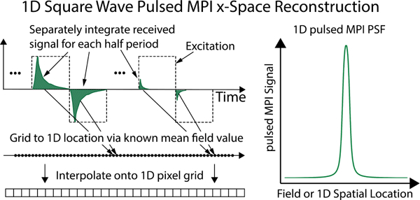
Fig. 5. Square wave data acquisition and image reconstruction using the AWR.

We integrate the signal for each square wave half-period and grid this value to the mean total applied field value. A slowly-varying bias field allows us to cover a large magnetic FOV. In this manner, we obtain a 1D square wave point-spread function (PSF). If we divide the applied field by a gradient strength (e.g., of a specific scanner) we can map to a 1D spatial PSF.
