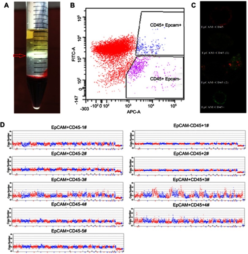
Figure 1.
Sample processing and CTC detection. Blood mononuclear cells (BMCs) and CTCs were isolated by density gradient centrifugation (A). EpCAM+ and CD45- cells identified by flow cytometry were considered tumor cells (B). Cells from different regions were identified by laser confocal microscopy (EpCAM-APC: Red: CD45-Alexa Fluor® 488: Green) (C). Cells randomly isolated by flow cytometry from the EpCAM+ CD45- region and the EpCAM+CD45+ region of one specimen were manually separated by micropipetting under a microscope, after which individual cells were subjected to whole-genome amplification to detect copy number variation (CNV) profiles. The copy numbers are segmented (blue and red lines) (D).

