
Figure 3.
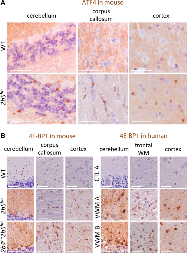
White and gray matter astrocytes in VWM mice and VWM patients are immunoreactive for ATF4‐regulated 4E‐BP1. (A) ATF4 immunoreactive nuclei (brown) are detected in white and gray matter macroglia of VWM mice. Two sections from brain tissue from 4‐month‐old WT and 2b5ho mice (n = 2) were stained with antibodies against ATF4. Findings in cerebellum, corpus callosum, and cortex are indicated. Staining for ATF4 on human brain sections was not successful. (B) White and gray matter astrocytes in VWM mouse and VWM human brain in show 4E‐BP1 immunoreactivity (brown). Two brain sections from 4‐month‐old mice (WT, 2b5ho, and 2b4he2b5ho), human control (C1) or patients (VWM 1 and VWM 2) were stained with antibodies against 4E‐BP1. Details on human brain tissue are listed in Data S5. Findings in cerebellum, corpus callosum (mouse) or frontal white matter (WM, human) and cortex are shown. Purple stain indicates nuclei. White bar, 0.05 mm.