
Figure 6.
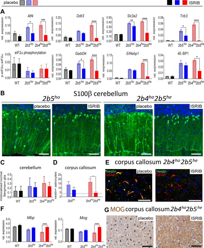
ISRIB modulates aberrant expression of ATF4‐regulated mRNAs and ameliorates neuropathological astrocyte markers in VWM mouse cerebellum and corpus callosum. (A) Cerebellar expression of ATF4‐regulated mRNAs, eIF2α phosphorylation, and 4E‐BP1 protein expression were measured in placebo‐ and ISRIB‐treated WT and VWM mice (n = 6 per group). Graphs show average ± SD. Two‐way ANOVA with Tukey’s correction was performed for each target. (B) Brain sections from placebo‐ and ISRIB‐treated 2b5ho and 2b4ho2b5he mice (n = 2 per group, 2 sections per animal) were stained with antibodies against S100β. Thickness of Bergmann glia (BG) processes is reduced by ISRIB in 2b4ho2b5he mice; white bar, 0.05 mm. (C) Counts of mislocalized and normally localized S100β‐positive Bergmann glia shows that ISRIB does not fully normalize Bergmann glia location in VWM mice. Differences in the ratio of mislocalized:normal localized Bergmann glia were statistically assessed by two‐way ANOVA with Tukey’s correction. (D and E) ISRIB reduces number of nestin‐GFAP double positive astrocytes in 2b4ho2b5he mice (2 sections per rostrum and splenium of the corpus callosum for 2 animals per group). The average number of nestin‐GFAP double positive astrocytes in four untreated WT animals was included as reference (indicated as dotted line in the graph). Statistical differences were determined with one‐way ANOVA using Tukey’s correction. White bar in immunofluorescence, 0.05 mm. (F) ISRIB restored levels of mature myelin mRNA markers in 2b4ho2b5he mice. Graphs show average ± SD (Akt + Gapdh, qPCR reference). Two‐way ANOVA was performed for each target using Tukey’s correction. (G) ISRIB restores immunoreactivity for mature myelin protein MOG in white matter structures in 2b4ho2b5he mice. Shown are representative sections from a total of four sections (n = 2 mice per group). Black bar, 0.05 mm. For all graphs, *P < 0.05, **P < 0.01, ***P < 0.001, ****P < 0.0001. Statistical test outcomes are in Data S6.