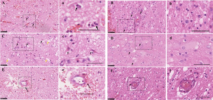
Figure 2.

Acute cellular changes following spontaneous ICH in human. (A) Presence of neutrophils (arrows), (a) and at higher magnification. (B) Recruited macrophages, (b) and at higher magnification. (C) Red Neurons (black arrows) and vacuoles demonstrating edema (yellow arrows), (c) and at higher magnification. (D) Reactive astrocytes (white arrows), (d) and at higher magnification. (E) A small vessel with erythrocytes dissecting its wall (arrow), (e) and at higher magnification. (F) Thickened small vessel endothelial cells (arrow), (f) and at higher magnification. Scale bar 50 µm. ICH, intracerebral hemorrhage.
