Figure 2.

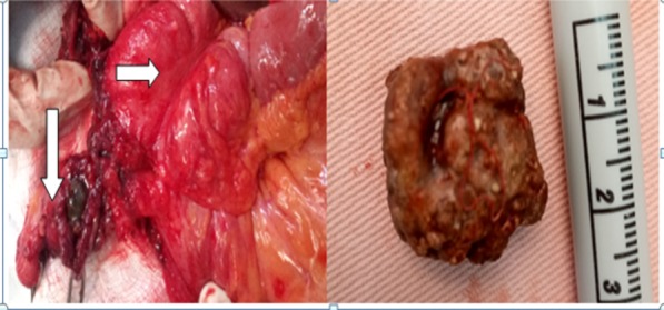
Left: intra-operative pictures, the perforated appendix with surrounding inflammatory changes, the stone inside (down arrow) and the Cecum (right side arrow), Right: The stone outside the appendix

Left: intra-operative pictures, the perforated appendix with surrounding inflammatory changes, the stone inside (down arrow) and the Cecum (right side arrow), Right: The stone outside the appendix