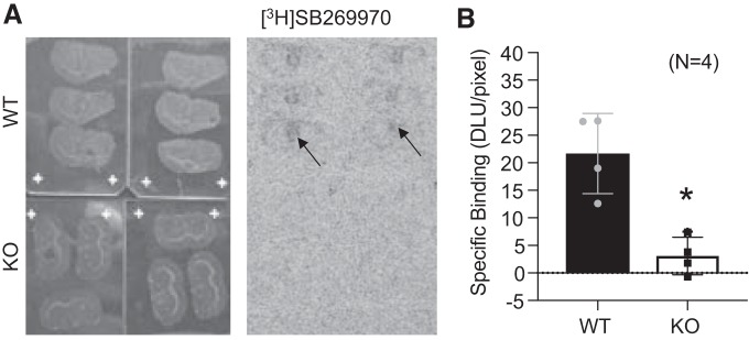
Fig. 7.
[3H]SB269970 radioligand binding site is lost in the 5-HT7 KO rat. Representative brain slides and correlated autoradiogram (A) and quantitation of the thalamus as a region of interest (arrows) for [3H]SB269970-specific binding in brain sections of the WT and KO rat (B). Bars represent means ± SE for number of points scattered around the mean. Four different rats, including male and female, are represented in this measure. An unpaired t-test was used to compare values of WT and KO. *P > 0.05 in all comparisons between WT and 5-HT7 KO tissues. DLU, digital light units.

