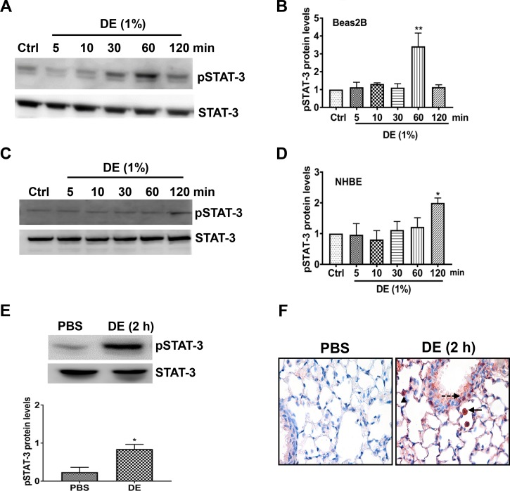
Fig. 2.
Dust extract (DE) induces STAT-3 activation in human bronchial epithelial cells and in mice. Beas2B (A and B) and normal human bronchial epithelial (NHBE, C and D) cells were treated with medium [control (Ctrl)] or 1% DE for the indicated times, and levels of pSTAT-3 and total STAT-3 were determined by Western blot analysis. Levels of pSTAT-3 were normalized to total STAT-3 levels. Normalized pSTAT-3 in control cells was arbitrarily considered as 1, and relative levels in treated cells are shown. Data shown are means ± SE (n = 4 for Beas2B, except n = 3 for 120-min treatment; n = 5 for NHBE). *P < 0.05, **P < 0.01 compared with cells treated with medium alone, according to one-way analysis of variance using Tukey’s multiple-comparison test. E: mice were administered 50 μl PBS or 50 μl 20% DE via intranasal administration, and 2 h later, pSTAT-3 and STAT-3 levels in lung homogenates were determined by Western blot analysis. Levels of pSTAT-3 were normalized to total STAT-3 levels, and relative levels in DE-treated mice are shown. Data shown are means ± SE (n = 4). *P < 0.05 compared with mice treated with PBS according to paired t-test. F: immunohistochemical detection of pSTAT-3 levels in lung sections of mice treated with PBS or 20% DE is shown. Arrow, inflammatory cell; dashed arrow, bronchiolar epithelial cell; arrowhead, alveolar type II cell. Magnification, ×400.

