Fig. 7.
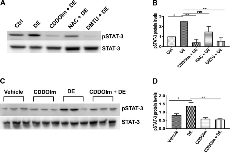
Antioxidants suppress STAT-3 activation. A and B: Beas2B cells were first incubated with medium [control (Ctrl)] or medium containing 0.5 µM 1-(2-cyano-3, 12, 28-trioxooleana-1, 9(11)-dien-28yl)-1H-imidazole (CDDOIm) or 15 mM N-acetylcysteine (NAC) for 3 h or 30 mM dimethylthiourea (DMTU) for 1 h and then treated with or without 1% dust extract (DE) for 1 h. Levels of pSTAT-3 and STAT-3 were determined by Western blot analysis, and pSTAT-3 level was normalized to STAT-3. Levels of pSTAT-3 in control cells were arbitrarily considered as 1, and relative levels in treated cells are shown. Data shown are means ± SE (n = 4). *P < 0.05, **P < 0.01; ns, not significant, according to one-way analysis of variance with Tukey’s multiple-comparison test. C and D: female C57BL/6 mice (n = 3) received vehicle or CDDOIm (5 mg/kg) once daily for 2 days via intraperitoneal injections before intranasal administration of 50 μl of PBS or 20% DE. After 2 h, mice were euthanized, and lung homogenates were prepared. Levels of pSTAT-3 and STAT-3 were determined by Western blot analysis, and pSTAT-3 level was normalized to STAT-3. Data shown are means ± SE. *P < 0.05, **P < 0.01 according to one-way analysis of variance using Tukey’s multiple-comparison test.

