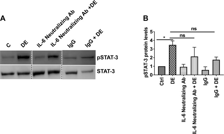
Fig. 10.
IL-6 is not solely responsible for STAT-3 activation. A and B: Beas2B cells were treated with 1 µg/ml of anti-IL-6 neutralizing antibody or matching control antibody for 1 h and then treated with 1% dust extract (DE) for 1 h. Levels of pSTAT-3 and total STAT-3 were determined by Western blot analysis, and pSTAT-3 level was normalized to total STAT-3. Dashed lines show reassembly of noncontiguous lanes. pSTAT-3 level in control (C/Ctrl) cells was arbitrarily considered as 1, and relative levels in other samples are shown. Data shown are means ± SE (n = 5); ns, not significant, *P < 0.05 according to one-way analysis of variance using Tukey’s multiple-comparison test.

