Fig. 5.
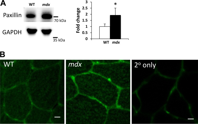
The focal adhesion protein paxillin is increased in mdx muscles in vivo. Focal adhesions regulate force transfer between the myofiber cytoskeleton and its environment. A: Western blots of paxillin and GAPDH from WT and mdx muscles. The cropped blots in each panel are from a single gel and single exposure of 2 contiguous lanes. Densitometry measures were normalized to GAPDH expression and to WT muscles. B: representative confocal cross sections showing paxillin labeling (green) in WT and mdx muscle sections. Labeling with secondary antibody only shows a low background signal. These methods show that mdx muscles have increased paxillin expression and labeling compared with WT muscles. Values are means ± SD for TA muscles from 3 animals/genotype. A t-test was performed on log-transformed data to determine statistical differences between groups. Scale bar = 10 μm. *P < 0.05 compared with WT.

