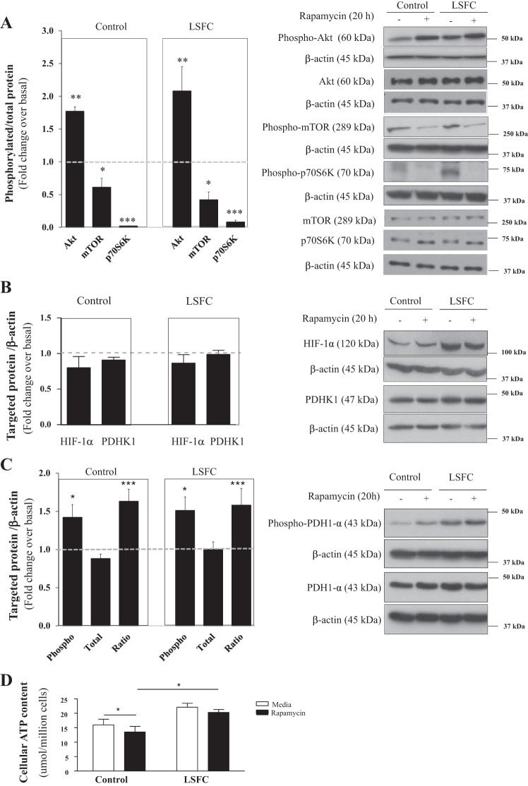
Fig. 4.
Suppression of mechanistic target of rapamycin (mTOR) complex 1 (mTORC1) activity does not alter hypoxia-inducible factor 1α (HIF-1α) and pyruvate dehydrogenase kinase 1 (PDHK1) levels. Primary fibroblasts from control and Leigh syndrome French Canadian type (LSFC) donors were treated for 16 h with 100 nM rapamycin or vehicle (DMSO) in supplemented DMEM. On the day of the study, cells were cultured in a serum-free nonsupplemented DMEM, with inhibitor or vehicle as indicated, for an additional 4 h. A: suppression of mTORC1 activity was confirmed by immunoblot and densitometric analysis of phosphorylated levels of mTOR (n = 3), p70 ribosomal S6 kinase (p70S6K, n = 3), and Akt (n = 3). B and C: immunoblot and densitometric analysis of total levels of HIF-1α (n = 7) and PDHK1 (n = 8; B) and phosphorylated pyruvate dehydrogenase 1α (PDH1-α, n = 7; C) in response to rapamycin treatment. D: primary fibroblasts were cultured with inhibitor or vehicle, as indicated, in opaque 96-well plates at a density of 10,000 cells per well in triplicate. Cellular ATP levels were measured with the ATPlite luminescence ATP detection assay system (n = 4). Results represent means ± SE. Difference between baseline and rapamycin treatment was assessed with a paired Student’s t-test. *P ≤ 0.05; **P ≤ 0.01; ***P ≤ 0.001.

