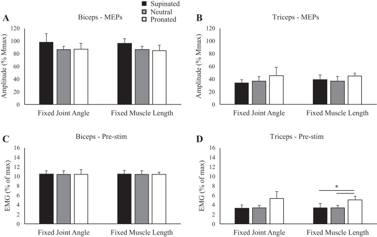
Fig. 5.
Group data (means ± SE, n = 10) during isometric elbow flexion for motor evoked potential (MEP) amplitudes of the biceps (A) and triceps brachii (B) and prestimulus (Pre-stim) electromyography (EMG) before transcranial magnetic stimulation for the biceps (C) and triceps brachii (D). Black, gray, and white bars correspond to supinated, neutral, and pronated forearm postures, respectively. MEP amplitudes are shown relative to the maximum M wave (Mmax) taken during the same conditions. EMG is normalized to the maximum EMG found during muscle-specific maximal voluntary contractions. *P < 0.05 between forearm postures.

