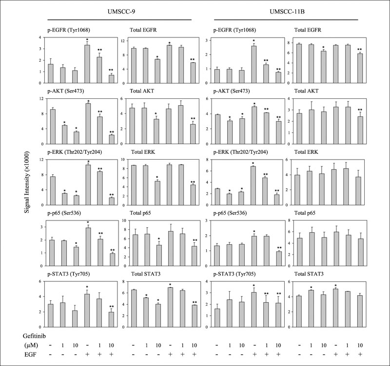
Fig. 5.
Reverse phase protein array detection of phosphrylated and total proteins in UMSCC cells lines. Reverse phase protein array was done using the same whole cell and nuclear extracts as used in the previous experiments. The images of immunohistochemical staining were scanned, and data were extracted by ImageQuant5.2 software from replicated slides. * indicates the statistical significance when compared with NoTx conditions; ** indicates the difference when compared with EGF-treated conditions (P < 0.05, Student’s t-test).

