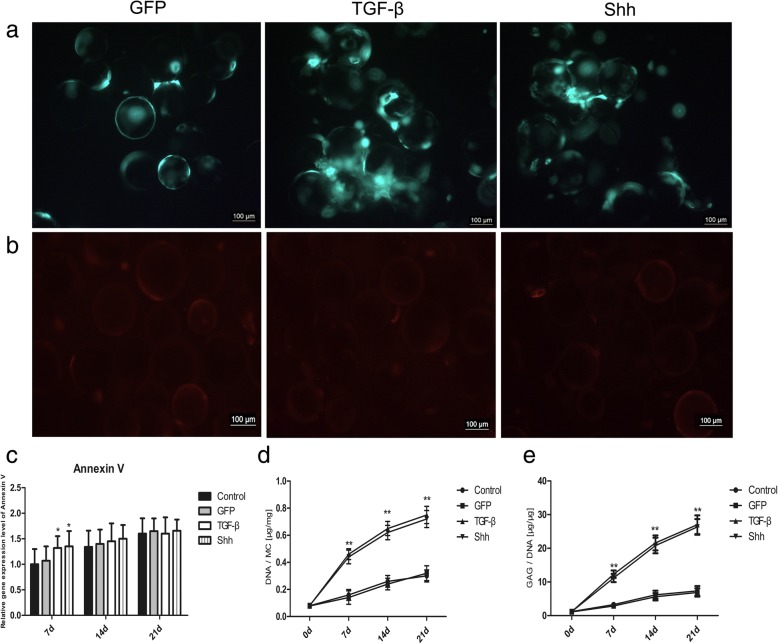
Fig. 3.
BMSCs viability and proliferation on cytodex 3. a BMSCs transfected with Shh or GFP viral plasmids revealed green fluorescence after 21 days of culture. GFP-transfected BMSCs treated with TGF-β served as a positive control. b Annexin V-PE immunofluorescence staining of BMSCs on cytodex 3 at day 21. Dead cells are labeled in red. c RT-PCR analysis of Annexin V on days 7,14 and 21 during differentiation induction. d, e Comparison of DNA and GAG/DNA ratio at different time points. MC, microcarrier. Significant differences between the control group are indicated by * p < 0.05, **p < 0.01

