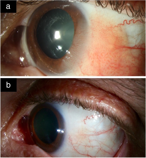
Fig. 4.

Slit lamp photo of OSSN before and after MMC treatment. a. Slit-lamp picture left eye with an extensive OSSN. Papillary fronds of this diffuse OSSN are noted on the bulbar, limbal and corneal surface. b. Slit-lamp picture after 3 weekly cycles of mitomycin-C (MMC) 0.04% with 2 to 3 weeks between cycles. The OSSN notably regressed with treatment
