Fig. 1.
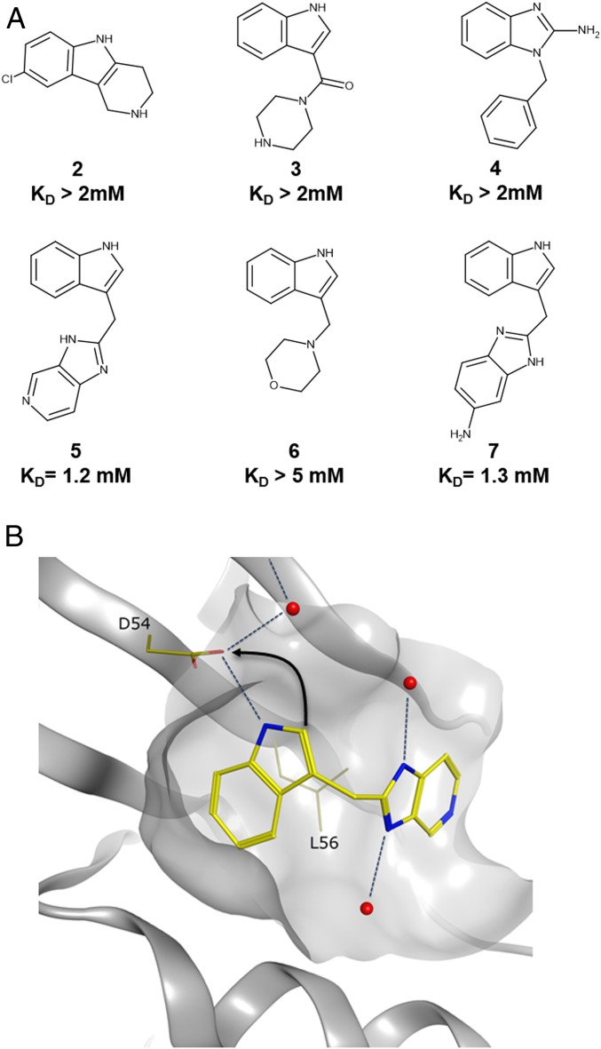
Fragments identified from 2 separate fragment screens. (A) Representative indole and benzimidazole fragments identified from the fragment screens. (B) The binding mode of indole 5 in GDP-KRAS (Protein Data Bank [PDB] ID code 4EPV) showing the H bond between the indole NH and the side chain of D54. Indole 5 shown in yellow, water molecules shown in red. Arrow indicates the strategy of forming an additional charge−charge interaction with the side chain of D54 from the indole 2 position.

