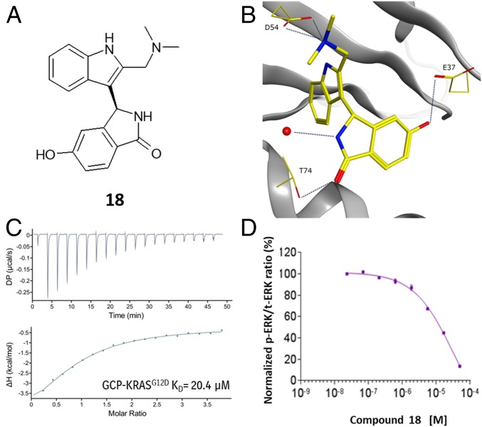
Fig. 3.
X-ray, biophysical and cellular data for 18. (A) Chemical structure of 18. (B) X-ray structure of 18 in GCP-KRASG12D, highlighting the polar interactions formed with D54, T74, and E37 (PDB ID code 6GJ6). (C) ITC dose titration curve for 18 and GCP-KRASG12D. (D) Meso Scale Discovery analysis of pERK levels in NCI-H358 cells after 2-h treatment of 18.

