Figure 3.
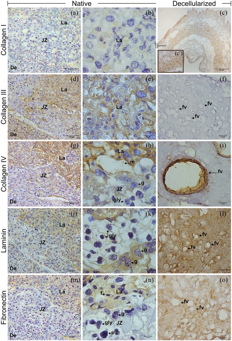
Immunohistochemistry for collagens I, III, and IV, laminin and fibronectin in native and decellularized mouse placenta. Collagen I was lightly visualized in native (a), (b) and unexpectedly strong spread in the ECM after decellularization (c), (c′). Unlike, the collagen III observed in native (d), (e), was absent in the scaffolds (f). Collagen IV (g), (h), laminin (j), (k), and fibronectin (m), (n) had similar distribution in the native placental regions, except by the fibronectin absence in the junctional zone (n). In the acellular ECM, the mainly collagen IV (i), but also laminin (l) and fibronectin (o) surrounded the former fetal vessels. De: decidua; JZ: junctional zone; La: labyrinth; f: fetal capillary; fv: former fetal vessel; g: trophoblast giant cell; gly: trophoblast glycogen cell; m: maternal blood space.
Scale bars = 100 µm (a, c, d, g, j, m), 50 µm (c′), 10 µm (b, e, f, h, i, k, l, n, o).

