Fig. 3.
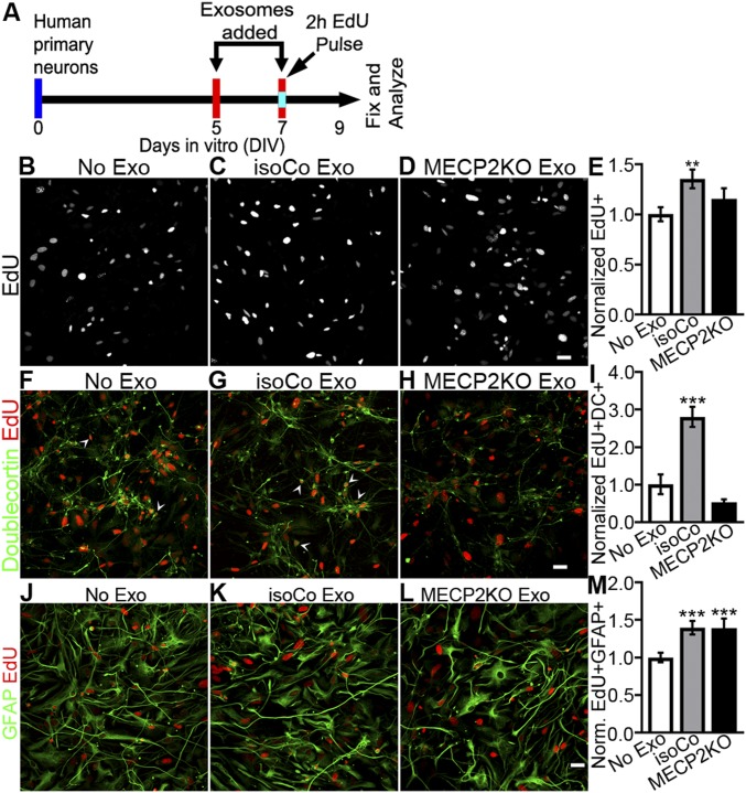
Isogenic control exosomes increase proliferation and neuronal fate in developing neural cultures, whereas MECP2LOF exosomes have no effect. (A) Protocol for treatment of human primary neural cultures with exosomes to assay proliferation, survival, and cell fate. Human primary neural cultures were treated with media alone or exosomes on DIV 5 and 7. On DIV 7, cultures were exposed to 10 µM EdU for 2 h just before the second exosome or media treatment. Cultures were fixed and immunolabeled for analysis on DIV 9. (B–E) Isogenic control exosomes increase cell proliferation. (B–D) Confocal images show EdU-labeled human neural cultures treated with media (B, No exo), isogenic control exosomes (C, IsoCo exosomes), and MECP2LOF exosomes (D). (E) Treatment with isogenic control exosomes (gray bars) increased EdU-labeled progeny by 1.35× ± 0.09 (P = 0.006) compared with no exosomes (white bar), whereas MECP2LOF exosome treatment (black bars) had no effect (E). Edu+ cell numbers normalized to no-media condition. (F–I) Isogenic control exosomes increase neuronal differentiation. Confocal images of EdU-labeled (red) and doublecortin-labeled (green) cultures treated with media (F), isogenic control exosomes (G), and MECP2LOF exosomes (H). Control exosomes (gray bars) increased neuronal progeny 2.8× ± 0.27 (P = 0.005), whereas MECP2LOF exosomes (black bars) had no effect (I). (J–M) Isogenic control exosomes and MECP2LOF exosomes increase astrocyte differentiation. Confocal images of EdU-labeled (red) and GFAP-labeled (green) cultures treated with media (J), isogenic control exosomes (K), and MECP2LOF exosomes (L). Control as well as MECP2LOF exosome treatments increase Edu+GFAP+ astroglial progeny by 1.4× ± 0.09 (P = 0.006) and 1.4× ± 0.13 (P = 0.009), respectively (M). n = 4 wells per group. Statistics computed with 2-way ANOVA with Bonferroni correction. (Scale bar, 20 µm.)

