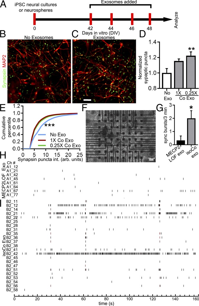
Fig. 5.
Control exosome treatment increases synapse density and neuronal firing in MECP2LOF hiPSC-derived neurospheres. (A) Protocol for treatment of MECP2LOF hiPSC-derived neural cultures or neurospheres with control exosomes or media alone to assay synapses and synchronized firing. (B–D) Control exosomes increased synapse density in MECP2LOF iPSC-derived neural cultures. Cultures were fixed on DIV 50 and labeled with the neuronal antibody MAP2 and Synapsin1 to identify synaptic puncta. Synaptic puncta density and intensity were quantified. Images of MAP2 (red) and Synapsin1 (green) labeling in MECP2LOF neural cultures without exosome treatment (B) or with control exosome treatment (C). (D) Exosome treatments increased synapse density. Treatment with control exosomes (0.25× dose) increased presynaptic puncta density to 1.22× ± 0.05 of control (no exosome) values (P = 0.006, n = 4 wells each, 2-way ANOVA with Bonferroni correction). (E) Kolmogorov–Smirnoff plot of cumulative frequency of synaptic puncta intensity shows that control exosome treatments (1× control [red line, D-stat = 0.24, P < 0.0001] and 0.25× control [green line, D-stat = 0.21, P < 0.0001]) increase the fraction of low-intensity puncta compared with no exosome treatment (blue line). (F–I) Treatment of MECP2LOF neurospheres with control exosomes increased neural circuit activity. MECP2LOF hiPSC-derived neurospheres were plated on 64-channel multielectrode array (MEA; F) and treated with MECP2LOF exosomes or control exosomes. (G) Graph showing that synchronized bursts of activity occur with a greater frequency in MECP2LOF neurospheres treated with isogenic control exosomes compared with neurospheres treated with MECP2LOF exosomes (MECP2LOF exo, 0.33 ± 0.33 bursts per 3 min; control exo, 2.0 ± 0.68 bursts per 3 min; P = 0.03, n = 3 arrays each, 2-tailed t test). (H and I) Aligned raster plots of spiking activity over 3 min of recordings from active channels. MECP2LOF neurospheres treated with isogenic control exosomes (I) have more overall activity and more synchronized activity across different electrodes compared with neurospheres treated with MECP2LOF exosomes (H). Note also the fewer active channels in neurospheres treated with MECP2LOF exosomes. (Scale bars: B and C, 50 µm; F, 100 µm.)

