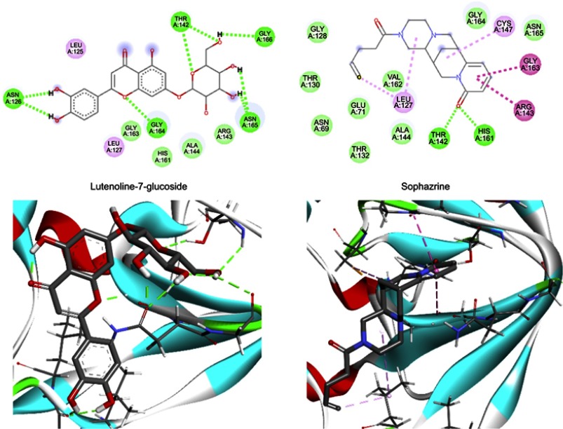
Figure 8.
The interactions between two top-scoring ligands and the amino acids of the active site of Coxsackievirus B4 3c protease (PDB ID: 2ZU3) depicted in 2D (top) and 3D (bottom). H-bonds, hydrophobic, and δ+-π stacking interactions are shown as green, light pink, and dark pink lines, respectively.
Abbreviation: PDB, protein data bank.

