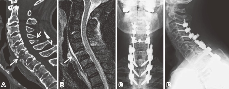
Figure 1.
(A) A sagittal reconstruction image of the CT scans showing ossification of the ALL, which was fractured along the C6 vertebral body, as part of the discovertebral three-column fracture throughout the C5 spinal process (arrows).
(B) A STIR MR image showing high signal in the C6 vertebra contagious to the prevertebral space; this image is consistent with hemorrhage in an extension injury (arrow).
(C) Anterior-posterior and (D) lateral radiograph status postposterior segmental screw instrumentation and autologous bone graft from C3 to T3 (C3-C5: lateral mass screws; T1-T3: pedicle screws).

