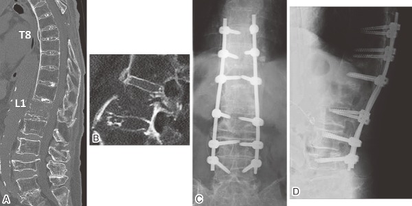
Figure 4.
(A) A CT scan image showing a healed T8 fracture status postlaminectomy and hardware removal.
(B) A magnified CT scan image showing a three-column extension-type fracture of the L1 vertebra.
(C) Anterior-posterior and (D) lateral radiograph status postpercutaneous segmental screw instrumentation from T10 to L4.

