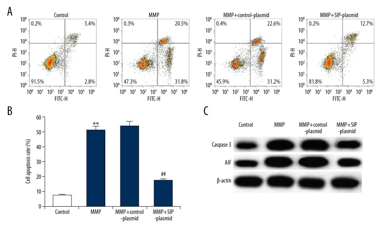
Figure 3.
Effect of SIP on MMP+ treated C6 cell apoptosis. C6 cells were transfected with control-plasmid and SIP-plasmid for 2 hours prior to treatment with 500 μM MPP+ for 24 hours, then cell apoptosis was analyzed by FCM (A); and the cell apoptosis rate was calculated and presented (B). The protein level of caspase-3 and AIF was detected using western blotting (C). Data were displayed as mean ±SD. ** P<0.01 versus control. ## P<0.01 versus MMP+ treatment alone group. SIP – steroid receptor coactivator-interacting protein; MPP+ – 1-methyl-4-phenylpyridinium; FCM – flow cytometer; AIF – apoptosis-inducing factor; SD – standard deviation.

