. 2018 Feb 27;5(1):63–74. doi: 10.3934/genet.2018.1.74

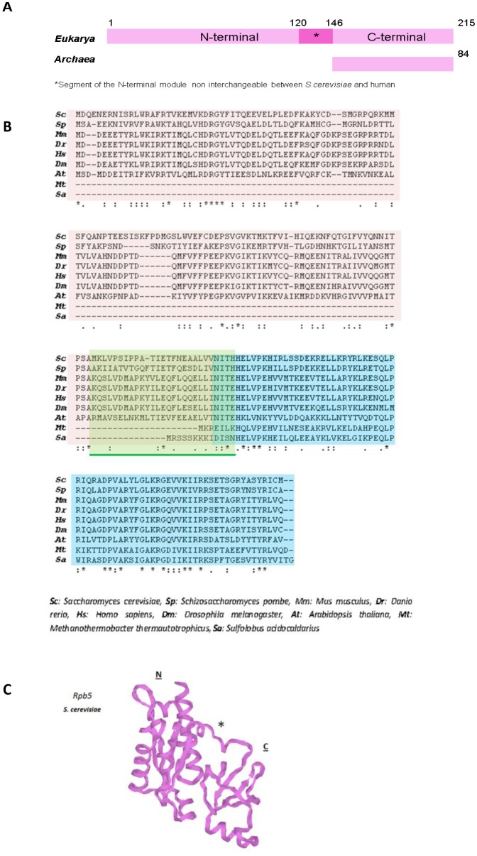
Figure 2. Comparison of Rpb5 and RpoH. (A) Schematic representation of eukaryotic Rpb5 and archaeal RpoH. The numbers on Rpb5 correspond to the amino acid positions in Saccharomyces cerevisiae and the numbers on RpoH to Sulfolobus acidocaldarius. (B) Comparison of Rpb5 and RpoH from different origins with clustal omega; *amino acid identity; “:” and “.” correspond to highly or less conserved amino acids, respectively. In clear brown, blue and green, the N-terminal, the C-terminal and the incompatibility domains, repectively. (C) Spatial organization of Rpb5 from S. cerevisiae (pdb: 1dzf). N: eukaryotic N-terminal domain (positions 1–142 in S. cerevisiae). C: C-terminal globe (positions 146–215 in S. cerevisiae). *Marks the segment of the N-terminal domain that is non interchangeable between S. cerevisiae and human.

Figure 2.