Figure 1.
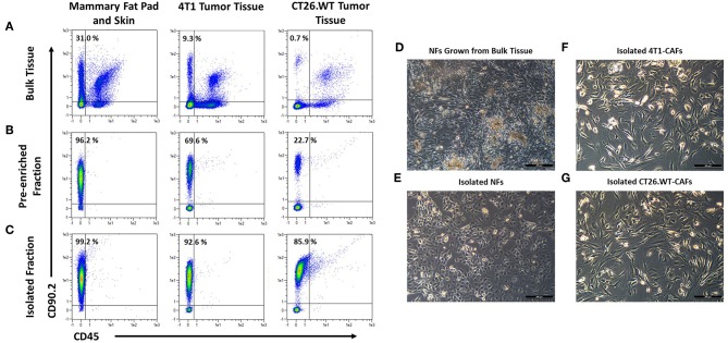
Isolation and cultivation of fibroblasts from solid tissues. (A–C) Flow cytometric analysis of cells before and after cell sorting. Fibroblasts, identified by expression of CD90.2 and absence of CD45, occurred at varying frequencies in different tissues (A). Fibroblasts were pre-enriched by depletion of CD90.2-expressing leukocytes and other non-fibroblast populations (B). Subsequently, CD90.2-expressing fibroblasts were labeled and magnetically separated from the pre-enriched fraction (C). (D–G) Images of cultivated fibroblasts. Normal fibroblasts, grown from single cell suspension of bulk tissues showed an elongated, spindle-shaped morphology, typical for activated fibroblasts (D), while fibroblasts isolated from healthy tissues showed the typical compact shape of normal fibroblasts (E). Isolated cancer-associated fibroblasts showed a spindle shaped morphology with an overall elongated appearance (F,G).

